
# INTRODUCTION:-
-> Centrioles are cytoplasmic organelles.
-> Mostly found in eukaryotic cells and are present in pairs called centrosomes.
-> Centriole lack a limiting membrane and DNA or RNA.
-> Forms the spindle of microtubules during cell division (mitosis and meiosis).
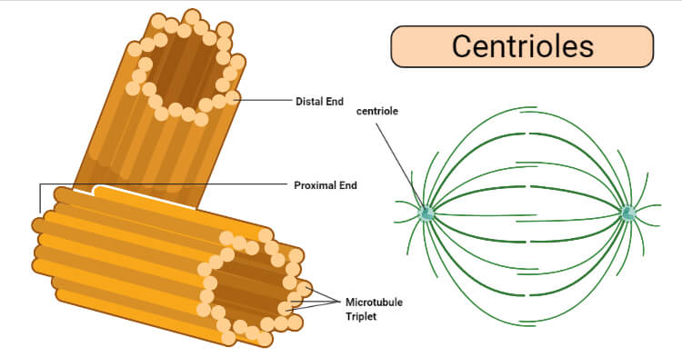
-> Each centriole lies at a right angle to the other.
-> Surrounded by amorphous pericentriolar material.
# STRUCTURE:-
-> The length of the centriole is about 3,000 to 5,000 Ã and the diameter is about 1,500 to 1,800 Ã.
-> Centrioles are made up of lipids and protein and also contain carbohydrates.
-> Centrioles are a set of microtubules. Arranged in the form of open-ended cylinders.
-> Made up mainly of tubulin protein (alpha tubulin and beta tubulin).
-> Centrioles mainly consist of 9 evenly spaced peripheral triplet fibrils of microtubules.
-> The nine triplet microtubules are arranged in a pattern of 9+3, linked with the help of an A-C linker.
# DUPLICATION:-
-> Before chromosome replication, the cell contains two centrioles(centrosome).
-> In the centrosome, the older centriole is termed the mother centriole and the other one is termed the daughter centriole.
-> During S-phase of the cell cycle, new centrioles grow at the proximal end of the mother and daughter centrioles.
-> The two pairs of centrioles remain attached to each other until mitosis.
-> They separate in mitosis with the help of the enzyme ‘separase’.
#FUNCTIONS:-
1) Cellular organisation;
-> Centrioles are involved in organising microtubules in the cytoplasm.
-> The position of the centriole in the cell determines the position of the nucleus and plays a critical role in the arrangement of cell organelles.
2) Formation of cilia and flagella:-
-> Centrioles form basal bodies, which give rise to axonemes of cilia and flagella.
3) In Cell division:-
∆ Mitosis.
• Mitosis is divided into four stages:-
1. Prophase
-> Prophase is marked by condensation of chromosomal material.
-> The nuclear membrane starts dissolving.
-> The centrioles(which are divided during S-phase) move to opposite ends of the nucleus.
-> The mitotic spindle fibres appear.
2. Metaphase
-> In metaphase, spindle fiber attached with the centromere at the kinetochore (a protein structure where the spindle fiber attached).
-> Centrioles are at opposite poles; centrioles pull the chromosomes to the centre of the cell (metaphase plate).
3. Anaphase
-> During anaphase, chromosomes split lengthwise, and sister chromatids separate.
-> The spindle fibres contract, and they pull one sister chromatid towards the opposite pole.
Tw4. Telophase
-> The cell division occurs in telophase.
-> The nuclear membrane reappears around the chromatid cluster.
-> The new daughter cells contain a pair of centrioles.
“From organising microtubules to helping cells divide, centrioles prove that even the smallest structures matter.”
Really amazing writter
ReplyDeleteyeye thank youuu
ReplyDelete